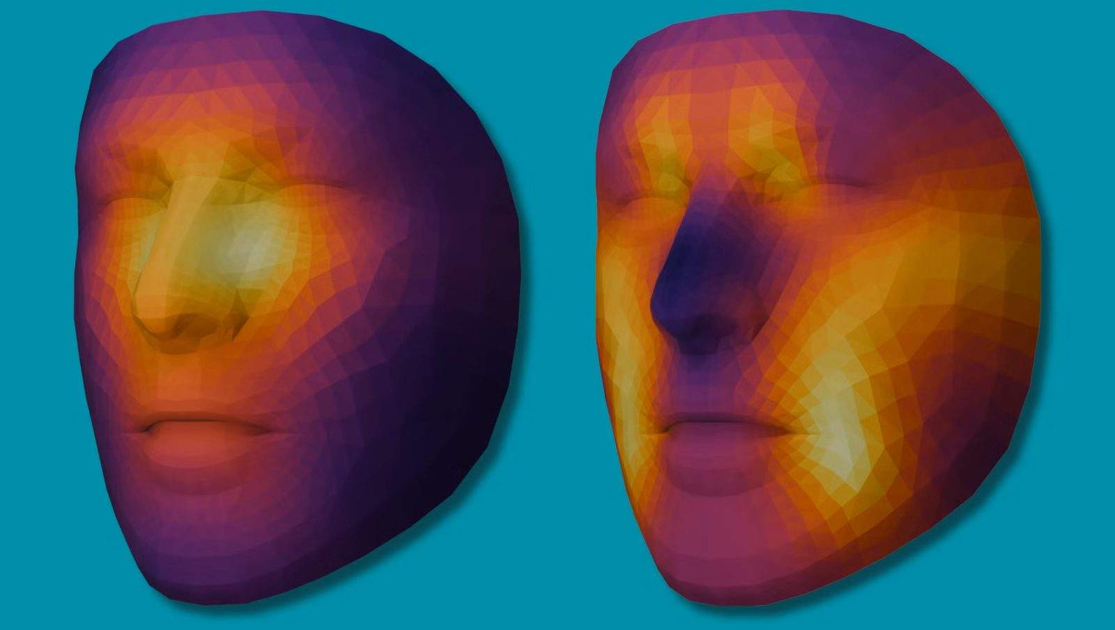
A single facial or body photograph cannot determine a patient's five-year survival rate on its own, yet computer vision models trained on visual phenotypes can extract systemic risk signals that standard intake forms miss. When researchers ask whether a photo can predict cancer survival, they are actually testing how reliably outward physical appearance correlates with biological frailty, disease burden, and treatment tolerance. This article maps that problem across four analytical dimensions: what visual signals actually represent biologically, why surface photography struggles as a standalone predictor, how multimodal data and bias controls determine real-world utility, and how clinical teams should deploy or reject image-based risk scoring.
The Signal Architecture Behind Visual Prognosis
Standard oncology prognosis relies on pathology reports, tumor staging, genomic profiles, blood biomarkers, and clinician-assigned performance scores such as Eastern Cooperative Oncology Group (ECOG) status. Each of these inputs measures a distinct slice of disease biology or functional capacity. Photographs sit in a different category: they capture high-dimensional, unstructured surface patterns that reflect downstream physiological effects rather than direct molecular drivers. You might also find this related story interesting: Why Women Are Calling Out Hidden Health Struggles Online.
In clinical practice, experienced physicians already read subtle visual cues during an exam. Pallor can point toward anemia or poor perfusion; temporal muscle wasting or prominent clavicles can reflect cancer-related cachexia; jaundice can signal hepatobiliary obstruction or hepatic involvement; periorbital edema or skin changes can accompany systemic stress, nutritional deficits, or treatment toxicity. A photograph freezes those external manifestations at a single point in time.
To treat a photograph analytically rather than metaphorically, it helps to separate the visual signal into three layers: As highlighted in latest coverage by Everyday Health, the results are significant.
- Morphological markers: Observable structural traits such as facial asymmetry, adipose distribution, muscle volume around the jaw and temples, skin texture, and visible lesions or surgical scars.
- Colorimetric and vascular cues: Subtle shifts in skin tone, conjunctival or mucosal redness, periorbital darkness, and peripheral perfusion that correlate with hemoglobin levels, hydration, oxygenation, or systemic inflammation.
- Behavioral and situational artifacts: Posture, head tilt, facial expression, fatigue indicators, or hospital lighting and clothing that often co-occur with severe illness even when they are not intrinsic tissue properties.
Distinguishing these layers matters because a neural network trained on raw images does not automatically know which pixels represent biology and which pixels represent the room around the patient. If a model scores a patient as high risk because hospital fluorescent lighting or a clinical gown appears in the frame, it has learned an environmental shortcut rather than a biological prognosis.
A known fact in oncology is that physical decline—especially involuntary weight loss, sarcopenia, and declining functional status—is strongly associated with worse survival across many malignancies and higher rates of treatment toxicity. An educated hypothesis in visual computing is that deep learning models can quantify subtle gradients of wasting, pallor, and physiological strain earlier or more consistently on a standardized image than a rushed clinician rating a subjective scale. Yet correlation in a retrospective dataset is not the same as causal prediction of survival. Cancer survival depends heavily on tumor histology, stage at diagnosis, molecular targets, access to timely therapy, and treatment response—variables a surface photograph cannot directly observe.
Why Surface Pixels Struggle as a Standalone Predictor
The central flaw in treating photo-based survival prediction as a direct substitute for clinical staging is information asymmetry. A photograph is a two-dimensional projection of external surfaces taken at one moment. Metastatic spread inside the liver, peritoneum, bone marrow, or central nervous system leaves no direct, unambiguous optical signature on a portrait. Two patients with identical facial appearance and body habitus can have vastly different prognoses: one may have early-stage, curable disease caught incidentally, while another has widespread occult metastasis that has not yet altered facial fat or skin tone.
Consider two patients presenting at an outpatient clinic with the same primary diagnosis. Patient A has localized disease and looks exhausted because of poor sleep and severe anxiety over a recent biopsy. Patient B has early systemic progression that has not yet caused visible weight loss or pallor, yet carries a much higher risk of relapse over the next three years. A photo-only model has no reliable way to separate anxiety-driven fatigue from indolent biology, nor can it see microscopic tumor burden inside internal organs.
The second limitation involves temporal dynamics. Cancer survival is a trajectory over months or years, whereas a photograph is a static snapshot. A single image cannot show whether a patient lost eight percent of their body weight over six weeks or has naturally been lean for twenty years. Without baseline comparison or longitudinal tracking, cross-sectional appearance conflates constitutional build with active catabolic decline.
Camera and capture conditions introduce another major failure mode. In real-world clinics and consumer settings, images vary across ambient lighting, white balance, camera sensor quality, lens distortion, makeup, facial hair, grooming, and pose. A model trained on well-lit clinical portraits taken against a neutral backdrop can suffer sharp performance drops when evaluated on smartphone selfies taken in dim rooms. Even minor shifts in color temperature can alter perceived pallor or skin tone, shifting predicted risk scores without any change in the patient's health.
There is also a subtle risk of circularity in model evaluation. If a study trains a model to predict survival using images collected from a tertiary cancer center, the model may absorb institutional selection effects—such as which patients were photographed, which stage groups were overrepresented, or which treatments were available during the study window. High accuracy in an internal test split does not guarantee generalizability to community clinics, different demographic populations, or newer treatment regimens that change survival curves altogether.
Multimodal Integration and Confounding Controls
Because surface images lack internal staging and longitudinal context, the highest-value role for visual data is as an auxiliary signal inside a multimodal risk model rather than a standalone oracle. When structured electronic health record variables, laboratory values, imaging reports, and pathology are combined with quantified visual features, the system can test whether the image adds incremental predictive lift beyond established clinical baselines.
Evaluating whether a photo genuinely adds value requires strict benchmarking. A model must beat a strong clinical baseline built from variables already routinely collected—such as age, cancer stage, histology, performance status, albumin, hemoglobin, and weight history. If adding image embeddings produces negligible improvement in discrimination or calibration once standard clinical variables are included, the visual pipeline adds operational complexity without clinical payoff.
Researchers and engineers building or auditing these systems must control for several high-impact confounders before claiming prognostic power:
- Demographic and skin-tone equity: Underrepresentation of darker skin tones or specific age and sex groups in training sets can distort colorimetric features and bias risk calibration across populations.
- Care-setting and socioeconomic proxies: Hospital background cues, clothing, image resolution, and capture timing often correlate with care access, comorbidity burden, or disease acuity at presentation.
- Treatment effect modification: Survival outcomes reflect both baseline disease biology and the therapies delivered afterward; a model trained on historical cohorts may misread risk if a newer targeted therapy or immunotherapy substantially improves prognosis for a subgroup.
- Label leakage and cohort censoring: Inconsistent follow-up windows, loss to follow-up, or non-cancer mortality can skew survival labels if time-to-event modeling is handled carelessly.
Calibration is just as important as ranking accuracy. Ranking two patients by relative risk is useful for triage, but clinical decision-making depends on whether a predicted probability aligns with actual observed survival rates in a given population. A model that ranks patients well on average can still overpredict risk in healthier outpatient cohorts or underpredict risk in frail inpatient populations.
Privacy and governance add another layer of constraint. Facial and body photographs are sensitive biometric identifiers that cannot be easily de-identified through simple metadata stripping. Re-identification risks, consent boundaries, storage security, and patient discomfort with being photographed during vulnerable moments can derail deployment even when algorithmic metrics look promising on paper.
Operationalizing Image Risk Models in Care Workflows
Even if a multimodal model demonstrates strong, unbiased predictive performance in validation studies, deploying it into real clinical workflows fails when teams treat risk scores as instructions rather than decision support. A risk estimate derived in part from appearance does not tell a clinician why a patient looks frail or which intervention will extend survival.
For oncology leaders, health-tech product managers, and clinical researchers evaluating photo-based survival tools, execution follows a disciplined sequence:
- Define the exact clinical decision point: Specify whether the score is meant to flag patients for early geriatric or nutritional assessment, stratify trial eligibility, prompt closer symptom monitoring, or support palliative care timing. A vague goal like "predict survival" leads to tools that nobody acts on.
- Establish a strict tabular baseline first: Build and validate a risk model using existing structured clinical and laboratory data, then measure the exact incremental gain—and calibration shift—from adding visual features across external cohorts.
- Audit subgroup error and confounding features: Test performance separately across age brackets, sexes, skin tones, disease stages, and image capture environments, and inspect whether background or lighting artifacts drive predictions.
- Enforce human-in-the-loop guardrails: Present risk outputs alongside transparent clinical drivers and uncertainty bounds, requiring clinician judgment and prohibiting automated treatment withholding or escalation based on a photograph alone.
- Monitor drift after deployment: Track input image quality, missingness, demographic shifts, and calibration over time as cameras, care settings, and oncology treatments evolve.
There are no silver bullets in prognostic modeling. A photograph cannot replace biopsy pathology, cross-sectional scans, or molecular diagnostics, nor can a model predict an individual's exact fate from a single picture. Treating visual risk scoring as an early warning flag for hidden physiological vulnerability—paired with rigorous external validation and clinical oversight—turns a promising computer vision concept into a practical check on blind spots in routine care.
The decisive strategic move for health systems and AI developers over the next three years is to stop pitching photo-based survival models as standalone prognostic tools and instead build them strictly as low-friction frailty and nutritional triage signals integrated into multimodal oncology workflows, gated by prospective external validation and subgroup calibration audits before clinical use.